- FRET의 한계 극복해 광유전학으로 조절가능한 바이오센서 개발···강력한 이미징 기술 될 것 -
암세포의 이동이나 신경세포가 활성화되는 장면을 관찰할 수 있다면 어떨까요? 세포의 다양한 변화를 포착하기 위해선 먼저 신호전달 스위치 단백질의 기능 연구가 먼저 필요합니다. 세포의 신호전달 스위치 단백질은 스위치가 켜지면 기계가 작동하듯 활성화 여부로 세포 기능을 제어합니다. IBS 연구진이 변화무쌍한 신호전달 스위치 단백질을 실시간 관찰할 수 있는 바이오센서를 개발하는데 성공했습니다.
우리원 인지 및 사회성 연구단(단장 신희섭, 이창준) 허원도 교수 연구팀은 대표적인 신호전달 스위치 단백질인 small GTPase 활성의 모든 변화 과정을 볼 수 있는 바이오센서를 개발했습니다. small GTPase 단백질은 세포의 이동, 분열, 사멸과 유전자 발현 등에 관여합니다. 세포의 핵심기능을 제어하기에 핵심 연구주제이기도 합니다.

small GTPase의 활성을 관찰하는 데엔 형광 공명 에너지전달(FRET)1)방식을 이용했습니다. 하지만 FRET 방식은 광유전학과 광 파장이 겹쳐 정작 관찰해야 할 세포신호의 변화는 보기가 어려웠습니다. 또 민감도가 낮아 동물 모델에 적용하는 것도 제한적이었습니다.
허원도 교수 연구팀이 그간 노하우를 바탕으로 개발한 바이오센서는 단백질 공학 기술로 5가지 종류의 small GTPase를 모두 관찰할 수 있습니다. 두 가지 파장(488nm, 561nm)을 활용하기 때문에 기존 바이오센서가 청색광을 활용하는 광유전학 기법의 파장과 겹치는 문제를 효과적으로 극복해 세포의 이동방향을 살피면서 동시에 공간적 기능도 분석할 수 있는 장점이 있습니다.
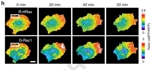
무엇보다 small GTPase 활성을 실시간으로 탐지할 수 있기에 암치료물질을 탐색하는 등 다방면의 기술 접목이 가능할 것으로 기대됩니다. 연구진은 실제 유방암 전이 암세포에 바이오센서를 발현시키고, 광유전학 기술로 암세포 이동 방향을 조절하자 small GTPase 단백질이 활성화됨을 확인했습니다. 이 과정에서 암세포의 이동 방향이 변할 때, 세포 내 small GTPase가 이리저리 움직이며 활성화하는 모습을 실시간 이미징하는데 성공했습니다.
더 나아가 IBS 연구진은 미국 막스 플랑크 플로리다 연구소(Max Plank Florida Institute)의 권형배 박사 연구팀과 공동연구를 진행했습니다. 연구진은 공 위를 달리는 실험으로 깨어있는 생쥐인 실험군과 마취된 대조군의 뇌 영역의 운동 피질2)의 신경세포에서의 small GTPase단백질의 활성을 비교하는데 성공했습니다. 살아있는 쥐에서 수 나노미터 단위의 신경세포 수상돌기 가시3)에서 실시간으로 변화하는 small GTPase 단백질의 활성을 관찰한 것은 이번이 처음입니다.

이번에 개발된 바이오센서는 시냅스처럼 수 마이크로미터 단위의 미세한 구조에서도 목표한 단백질을 관찰할 수 있을 만큼 민감도가 큽니다. 광유전학과 결합해 다양한 방식으로 관찰이 가능하고 민감도가 크기에 생체 내 두꺼운 조직 안에서 벌어지는 수 나노미터(nm) 크기의 변화까지도 정밀하게 볼 수 있습니다. 실험쥐의 운동행동과 같은 생리학적 현상에 지장을 주지 않는 자연스러운 상태에서 뇌 영역을 바로 실시간으로 관찰할 수 있어 뇌 관련 연구에도 다양하게 적용될 수 있습니다.
연구를 이끈 허원도 교수는 "이번 연구는 small GTPase 단백질을 생체 내에서 관찰하기 위한 기존의 바이오센서들의 기술적 한계를 극복하는데 성공했다"며 "특히 청색 빛을 활용한 광유전학 기술과 동시에 적용할 수 있어 다양한 세포막 수용체와 관련된 광범위한 세포신호전달연구와 뇌인지 과학연구에 접목이 가능할 것으로 기대된다"고 말했습니다.
연구결과는 세계적 학술지인 네이처 커뮤니케이션즈(Nature Communications, IF 12.353)에 1월 14일 오후 7시(한국시간) 온라인에 게재되었습니다.


1) FRET(Fluorescence Resonance Energy Transfer) : 두 가지 형광단백질간의 형광 세기변화의 비율을 측정하는 기술로 하나의 형광이 일으킬 수 있는 에너지가 다른 형광 물질에 전달되어 형광이 발현되는 현상을 이용한 관찰 기법이다.
2) 운동피질 (motor cortex) : 대뇌 반구에서 중심구 앞쪽 신피질 영역에 존재하며 몸의 움직임을 통제해 근육 운동에 관여하는 뇌 영역
3) 수상돌기 가시(dendritic spine) : 시냅스 후 구조인 수상돌기 가시는 다른 신경세포에서 신호를 전달 받는다.
