
Seminars & Events
Highlight
David Shum of Institut Pasteur Korea Introduces an Integrated Imaging and AI Platform for Infectious Disease Drug Discovery
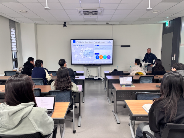
On November 17, the Center for Genome Engineering (CGE) at the Institute for Basic Science hosted David Shum, Head of the Screening Platform Lab at Institut Pasteur Korea (IPK), for an internal seminar titled “From Images to Insights: Integrative Platforms for Infectious Disease Therapeutics.”
Shum earned an M.S. in Biology from New York University and spent approximately a decade at the High-Throughput Screening Core Facility of Memorial Sloan Kettering Cancer Center, where he worked on assay development and large-scale screening projects. Since joining IPK in 2015, he has led the development of high-throughput and high-content screening platforms for viral, bacterial, and parasitic infection models.
The seminar introduced a drug discovery workflow that connects automated microscopy with large chemical libraries, molecular analysis, and artificial intelligence. At the center of this approach is the use of cellular images not merely as visual records but as high-dimensional data that quantify disease states and drug responses.
High-Content Imaging for Infectious Disease Drug Discovery
IPK’s screening infrastructure combines BSL-2+ and BSL-3 facilities with automated systems for liquid handling, compound dispensing, and detection. Its libraries range from FDA-approved and bioactive compounds to hundreds of thousands of small molecules and natural products. RNA interference platforms can also be used for focused or genome-scale target discovery.
In a typical high-content screen, cells are infected with a pathogen, exposed to candidate compounds at multiple concentrations, and imaged with automated microscopy. Unlike assays that measure a single endpoint, this approach can capture infection rate, cell number, morphology, and toxicity at the same time. It therefore helps identify active compounds while revealing unwanted effects on host cells.
The platform was also used during the COVID-19 pandemic to rapidly evaluate FDA-approved drugs for activity against SARS-CoV-2. Repurposing compounds with existing clinical information can be particularly valuable when a newly emerging pathogen leaves little time for conventional drug development.
Capturing the Morphological Signature of Chagas Disease with Cell Painting
Shum used Chagas disease as a case study in image-based morphological profiling. The disease is caused by infection with the parasite Trypanosoma cruzi. In the screening model, U-2 OS cells were infected with the parasite, treated with compounds, and cultured for 72 hours before imaging. An effective treatment can shift the morphology of infected cells toward that of uninfected cells, producing a measurable pattern of disease reversion.
Cell Painting uses multiple fluorescent dyes to label cellular structures including DNA, RNA, endoplasmic reticulum, mitochondria, F-actin, and the Golgi apparatus. Hundreds to thousands of morphological measurements can then be extracted from each cell. In the case presented, approximately 6,000 features were collected and reduced through quality control, missing-value handling, normalization, and feature selection.
Profiles from infected and uninfected cells were compared with those produced by the reference drugs benznidazole and posaconazole and by compounds with known mechanisms of action. Dimensionality-reduction methods such as UMAP and PCA revealed clusters of compounds with similar morphological responses. This made it possible to estimate both the extent of disease reversion and the likely mechanism of action of candidate compounds. Parallel tests without parasites helped separate host-cell activity and toxicity from antiparasitic effects.
Extending Morphological Profiling with AI
As image datasets grow, accurate cell segmentation and meaningful feature extraction become increasingly important. The team is developing a pipeline that combines Cellpose and the Segment Anything Model for segmentation, DINO-based vision transformers for feature extraction, and downstream mechanism-of-action classification. Dose-dependent profiles can further reveal whether a compound produces different biological effects as its concentration changes.
Future work will extend the platform to live-cell imaging, label-free analysis, and three-dimensional disease models. Organoids present a particular challenge because their cells are densely packed and structurally complex, requiring segmentation and profiling methods adapted beyond those used for two-dimensional monolayers.
The seminar demonstrated how large collections of cellular images can become a resource for evaluating efficacy, toxicity, and mechanism of action together. By integrating automated screening, cell biology, data science, and AI, this platform may help prioritize therapeutic candidates and accelerate the response to emerging infectious diseases.
References
Aulner, N., Danckaert, A., Ihm, J., Shum, D., & Shorte, S. L. (2019). Next-generation phenotypic screening in early drug discovery for infectious diseases. Trends in Parasitology, 35(7), 559–570.
Bray, M.-A., et al. (2016). Cell Painting, a high-content image-based assay for morphological profiling using multiplexed fluorescent dyes. Nature Protocols, 11(9), 1757–1774.
Jeon, S., et al. (2020). Identification of antiviral drug candidates against SARS-CoV-2 from FDA-approved drugs. Antimicrobial Agents and Chemotherapy, 64(7), e00819-20.

- 55, Expo-ro, Yuseong-gu, Daejeon, South Korea, 34126 Tel. +82-42-878-8310
Copyright(c) 2022 Center for Genome Engineering Research at IBS. All Rights Reserved.