Advanced Microscopy Facility
Super sensitive high resolution confocal laser scanning microscope for live cell
Dr. Jung Me Hwang Principal technician- + 82-52-217-5533
- hjm072@ibs.re.kr
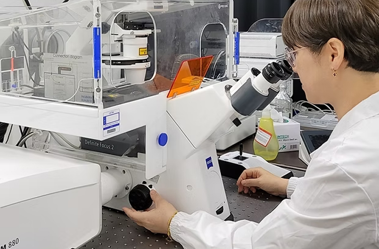
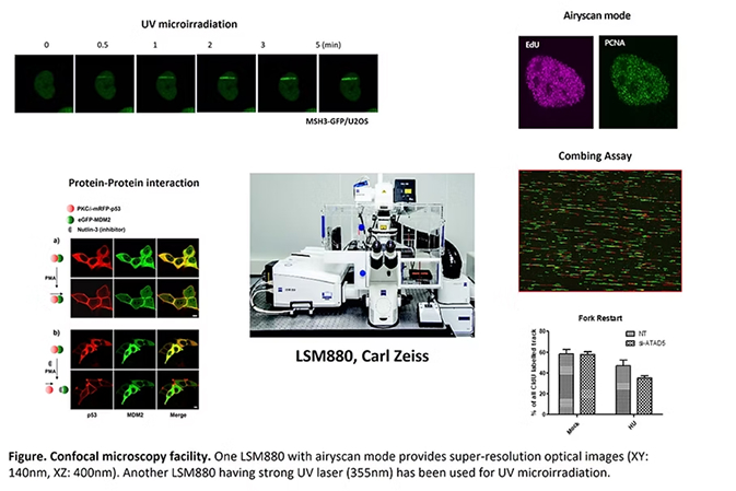
The confocal microscopy facility has two LSM880 confocal microscopes from Carl ZEISS. One LSM880 has an airyscan feature for improving the resolution of images. The facility provides high-resolution optical images with depth selectivity in three-dimensional reconstructions of complex objects. The facility provides education, assistance with data analysis, and maintenance of microscopes. The confocal microscopy facility has been extensively used for super-resolution of the live cell imaging, protein relocation in response to sites of DNA damage sites, scanning of DNA fibers to analyze DNA replication, fixed cell imaging, and protein-protein interactions. Results produced by the facility have been used in almost all publications from IBS-CGI.
Application- Super-Resolution Live Cell Imaging with standard multi-color live samples
- Super-Resolution at high penetration depth for large and thick samples
- UV Irradiation : for Detection of DNA recovery
- Tile Scanning : for experiment of DNA fiber / Combing assay
- Fixed Cell Imaging for Immunostaining & linear scanning
- Detection of cell cycle during long-term live cell
- Protein-Protein Interaction with CUPID system
- ROI-HDR, FRET, FRAP, FCS and 3D Visualization
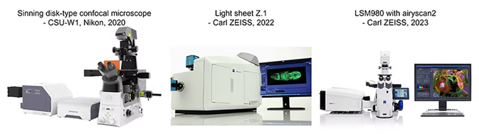
For time lapse imaging, in 2020 we also set up a Spinning Disk-Type Confocal Microscope that is capable of relatively low light delivery and the high spatial and temporal resolution, that cannot be achieved on our current laser scanning confocal or wide field microscopy systems. Based on testing equipment at Dundee and Edinburgh Universities (both Scotland), and also considering input from laboratories at the University of San Diego (USA) we set up a system provided by Nikon. This microscope which is also equipped such that FRAP and photoconversion allowing for analyzing the dynamic properties of proteins in vivo in real time can be conducted, is part of the C. elegans facility (see below).
In 2022, we get a ZEISS Light sheet Z.1 from the closed Plant aging life study group. The coverslip-free sample preparation for Lightsheet Z.1 and Multiview imaging give you the unique opportunity to view your sample from any angle. This microscope can imagine large samples like Zebrafish in 3D and long-term imaging of plants.
In 2023, we will set up a ZEISS LSM980 with Airyscan 2 that is capable of super resolution imaging , and is preparing for the disability of old confocal microscopy.

유전체 항상성 연구단
Room 224, UNIST Building 103, UNIST-Gil 50, Eonyang-eup, Ulju-gun, Ulsan, Republic of Korea
Copyright © Center for Genomic Integrity at IBS. All Rights Reserved.